Custom 3D-printed MRI sensors could transform imaging for children
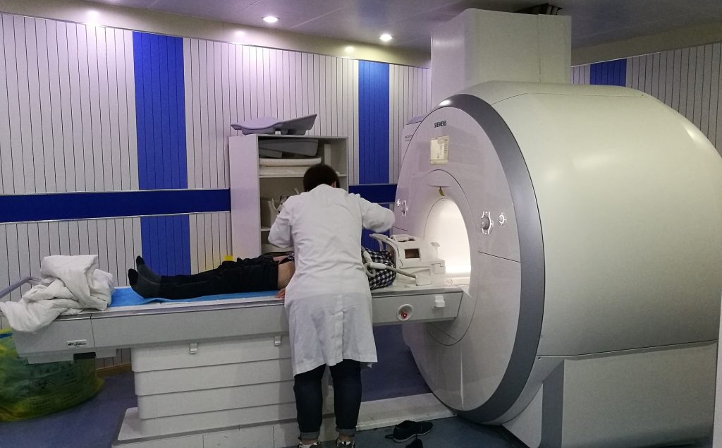
Medical imaging has revolutionised healthcare, enabling physicians to look inside the human body without invasive procedures. Yet many diagnostic tools are still designed around adult anatomy, creating challenges when imaging newborns, infants and young children. Researchers at the University of Southern California (USC) have now developed a promising solution: customisable, low-cost MRI sensors that can be 3D printed in minutes and tailored to individual patients.
The technology has the potential to improve image quality dramatically while reducing manufacturing costs and production times, opening new possibilities for paediatric medicine and personalised imaging.
The challenge of imaging small patients
Magnetic Resonance Imaging (MRI) remains one of the most powerful diagnostic tools available. By using magnetic fields and radio waves, MRI scanners generate highly detailed images of internal tissues and organs. However, image quality depends heavily on specialised devices known as coils, which act like antennas that receive signals emitted from the body.
For adults, standardised commercial coils generally perform well. For infants and children, the situation is more complicated. A baby’s heart, for example, may be only the size of a walnut. Commercial MRI coils are often too large, leaving gaps between the sensor and the patient’s body. These gaps reduce signal strength and image quality. The problem is further complicated by growth. A child may quickly outgrow equipment optimised for a particular body size. Custom imaging devices are available, but they can cost thousands of dollars and require lengthy manufacturing processes.
A new approach using additive manufacturing
Researchers led by Dr Yasser Khan, Assistant Professor of Electrical and Computer Engineering and Biomedical Engineering at the USC Viterbi School of Engineering, sought to redesign MRI coils from the ground up. Their solution combines flexible electronics with additive manufacturing. The team developed MRI sensors that can be customised digitally, printed in less than ten minutes and produced for approximately $30 per device. Most remarkably, testing showed that the customised sensors generated around four times greater image contrast than conventional commercial alternatives.
According to Khan: “By making customized MRI equipment faster and more affordable to produce, we have the potential to bring better imaging to patients who have traditionally had fewer options, especially infants and children.” The innovation is based on the concept of creating coils that conform closely to the patient’s anatomy, maximising signal capture and improving imaging performance.
MRI coils work best when positioned close to the area being examined. The further away the coil is from the target anatomy, the weaker the signal becomes.
Khan compares the issue to photography. “If you use a large lens to image something very small, you’re not going to get the clearest picture,” he explains. “But if you can tailor the lens, in this case the MRI coil, to the individual patient, you can capture a much better image.”
A customised coil that closely follows the contours of an infant’s chest, for example, can capture stronger signals from the heart than a generic adult-sized device.
The research team spent approximately three years evaluating different materials, polymers and conductive substances before arriving at the final design. Their solution involves printing conductive silver ink onto a stretchable thermoplastic elastomer, a material with properties similar to human skin. The resulting sensor can stretch by approximately 5 to 10 per cent while maintaining electrical performance. This flexibility allows the device to conform comfortably and securely to the body surface, even in areas where anatomy changes shape during movement or breathing.
The researchers also faced the challenge of integrating the printed sensors into existing MRI systems. This required designing specialised electronics capable of connecting the flexible coils to conventional imaging equipment. The outcome is a platform that can rapidly generate patient-specific devices. Rather than waiting months for a manufacturer to produce a customised coil, clinicians could potentially adjust a digital model and print a new version within minutes.
Imaging anatomy in motion
The USC project demonstrates the value of multidisciplinary collaboration. The research was enabled through cooperation between two groups at the USC Michelson Center for Convergent Bioscience. The DISC laboratory houses a unique MRI platform capable of imaging anatomy during motion, allowing researchers to study dynamic physiological processes that are difficult to capture using conventional MRI approaches.
This capability could prove particularly valuable for paediatric cardiology. Capturing clear images of a rapidly beating infant heart has traditionally been one of the most demanding applications in MRI.
John Wood, Director of Cardiovascular MRI at Children’s Hospital Los Angeles and Professor of Paediatrics and Radiology at the Keck School of Medicine of USC, is among the clinical collaborators helping assess where the technology may have the greatest impact.
Improved imaging quality could aid diagnosis, treatment planning and long-term monitoring of congenital heart conditions and other paediatric disorders. Beyond paediatrics, the technology highlights a broader trend in medicine: the movement towards patient-specific devices. Three-dimensional printing has already transformed areas such as prosthetics, surgical planning and medical implants. The USC system suggests that imaging devices may soon join that list.
The ability to rapidly fabricate highly customised MRI sensors could have applications for adult patients as well, particularly for unusual anatomies, rare conditions or specialised imaging procedures where conventional coils perform poorly. The work also demonstrates the power of bringing engineers, imaging scientists and clinicians together around a common challenge. As Khan notes: “This project wouldn’t have happened without access to the MRI and conversations with cardiologists, radiologists and imaging scientists. When you bring that expertise together, you can solve problems none of us could solve alone.”
Custom 3D-printed MRI sensors could transform imaging for children
#Custom #3Dprinted #MRI #sensors #transform #imaging #children