Q&A: Radiation-free 3D imaging boosts breast cancer screening
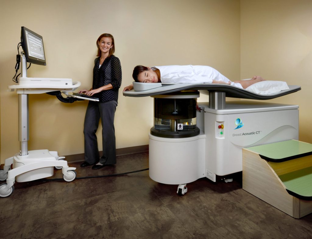
QT Imaging is a company advancing a quantitative, radiation-free, and compression-free 3D breast imaging platform across global markets. The company uses Breast Acoustic CT™, which deploys low-frequency transmission and reflection sound waves to generate true 3D, high-resolution anatomical maps without ionizing radiation or painful breast compression.
Digital Journal spoke with CEO Dr. Raluca Dinu about how the technology differs from traditional mammography.
Digital Journal: Could you start by sharing a bit about your background and providing an overview of QT Imaging’s mission?
Raluca Dinu: At QT Imaging, we are redefining what’s possible in breast imaging by delivering safe, high-resolution, and cost-effective solutions that address the limitations of today’s technologies, particularly for women with dense breasts, which affect approximately 50% of women in the United States and up to 75% in many Asian countries. Our mission is to bring the first quantitative, 3D, safe imaging platform to breast diagnostics and precision breast oncology. The future of breast imaging lies in providing physicians with richer biomarker-based diagnostic information, extracted from unique set of data, to support more informed clinical decisions, while offering women a safe, comfortable, and radiation-free imaging experience.
On a personal note, I hold a Ph.D. in Physics, and I thrive at the intersection of science, technology, and business. My passion is transforming breakthrough science into commercially successful solutions. As a turnaround executive, QT Imaging is a perfect fit because it combines innovative technology with the opportunity to build a company that can have a meaningful impact on women’s health. At the end of the day, it’s all about making a meaningful difference for patients.
DJ: What exactly is the Breast Acoustic CT Scanner, and how does your technology differentiate itself from traditional mammography or breast MRIs?
Dinu: QT Imaging’s Breast Acoustic CT Scanner is the first commercially available breast imaging system that uses sound waves, rather than ionizing radiation, to create 3D, high-resolution, quantitative images of the entire breast. Unlike mammography, it requires no painful breast compression or radiation. Unlike MRI, it requires no intravenous contrast agent and is designed to be a more cost-effective and accessible imaging solution.
Dense breast tissue is one of the greatest challenges in breast imaging because both dense tissue and cancer appear white on a mammogram, making cancers more difficult to detect. While MRI is highly sensitive in women with dense breasts and is often used as a supplemental imaging modality, it is expensive, requires intravenous contrast, has relatively low specificity, and is not practical for widespread screening.
QT Imaging’s technology was designed specifically to address these challenges. Rather than simply producing images, we quantitatively measure the physical properties of breast tissue using sound waves to generate objective, biomarker-based information. We believe this quantitative approach has the potential to improve tissue characterization, support more informed clinical decision-making, and advance precision breast oncology, while providing women with a safe, comfortable, and radiation-free imaging experience.
DJ: With U.S. FDA breast density disclosure mandates gaining traction, why is imaging dense breast tissue such a challenge for providers, and how does QT Imaging solve this?
Dinu: The FDA’s nationwide breast density notification requirement, which became effective in September 2024, reflects growing recognition that breast density is a major issue in women’s health. For the first time, every woman undergoing a mammogram in the United States must be informed if she has dense breast tissue because dense tissue not only increases the risk of breast cancer but also makes cancers more difficult to detect with mammography. As millions of women become aware of their breast density, the need for safe, effective, and accessible supplemental imaging technologies has never been greater. That’s exactly the unmet clinical need QT Imaging is designed to address.
QT Imaging addresses this challenge through a fundamentally different approach to breast imaging. Our technology captures both transmitted and reflected sound wave information, providing a spectrally rich dataset that extends well beyond conventional anatomical imaging. From these data, we derive quantitative biomarkers based on the physical properties of breast tissue, including the speed of sound, reflection intensity, attenuation, and other acoustic parameters. Together, these biomarkers provide objective information about tissue composition and heterogeneity, with the potential to improve tissue characterization and support more informed clinical decision-making, particularly in women with dense breasts.
DJ: What major trends are you seeing emerge right now in healthcare AI validation and quantitative tissue biomarkers?
Dinu: We’re seeing two important trends converge. The first is the rapid adoption of AI in radiology. AI is increasingly becoming an integral part of clinical practice, supporting everything from image acquisition and reconstruction to lesion detection, workflow optimization, and diagnostic decision support. As AI continues to mature, the emphasis is shifting from algorithm development to the quality of the underlying data. The future belongs to AI trained on rich, quantitative, clinically validated datasets that can generate reproducible and clinically meaningful results.
The second trend is the transition from one-size-fits-all medicine to personalized healthcare, driven by quantitative imaging. Clinicians are increasingly looking beyond anatomical images to objective, quantitative biomarkers that reflect the biological characteristics of an individual patient’s tissue. Rather than simply detecting abnormalities, the future of breast imaging is to measure them, providing reproducible biomarkers that can improve tissue characterization, assess disease biology, monitor response to therapy, and support more personalized treatment decisions. We believe this quantitative approach will be fundamental to precision breast oncology, where quantitative, objective information has the potential to help physicians select the most appropriate treatment for each patient, monitor disease safely over time, and, whenever clinically appropriate, support breast-conserving therapies.
DJ: What is your strategy for disrupting this space and convincing hospital systems to adopt a brand-new platform?
Dinu: Our strategy is to build clinical and economic evidence that demonstrates where our technology adds value within the existing breast imaging pathway. Adoption in healthcare is driven by evidence, not novelty.
That starts with rigorous clinical validation. We are collaborating with leading academic institutions and breast imaging specialists to generate evidence demonstrating where our technology can improve patient care, particularly for women with dense breasts and in precision breast oncology. Equally important, our platform is designed to integrate into existing radiology workflows rather than disrupt them.
Finally, we believe adoption will depend on delivering value to all stakeholders. Physicians need better diagnostic information, patients want a safer and more comfortable imaging experience, hospital systems need cost-effective technologies with clear clinical utility, and payers increasingly expect evidence of improved outcomes and value. Our strategy is to address all of those needs simultaneously.
Q&A: Radiation-free 3D imaging boosts breast cancer screening
#Radiationfree #imaging #boosts #breast #cancer #screening